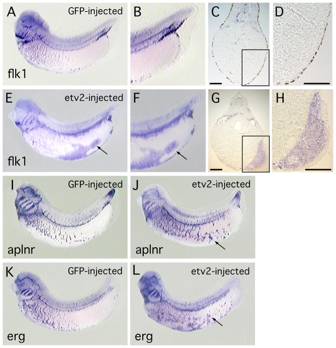
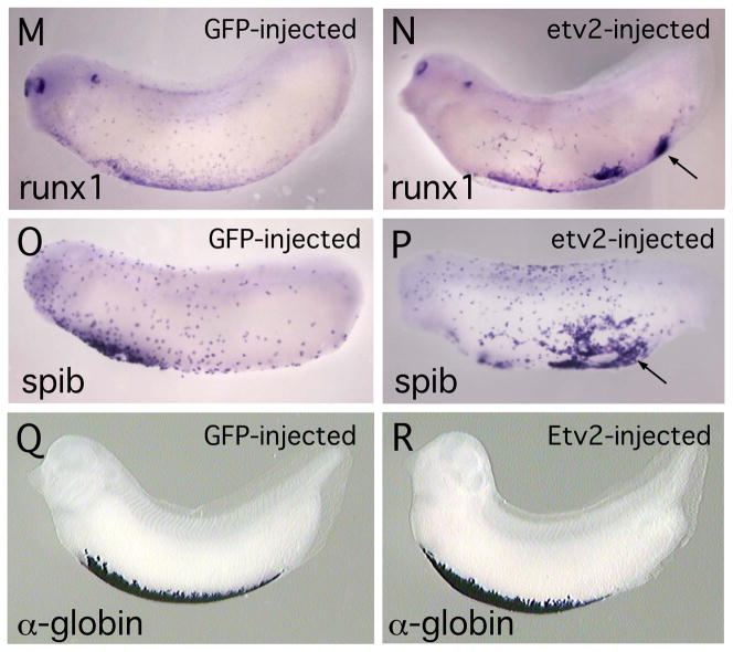
Fig. 3. Forced expression of ETV2 in the Xenopus embryos results in ectopic expression of endothelial and myeloid marker genes.
(A–D). Control embryo, injected with EGFP mRNA and assayed for flk1 transcripts. (A) View of whole embryo, and (B) enlarged view of ventral posterior region of embryo showing near absence of endothelial (flk1-expressing) cells. (C) Transverse section through embryo and (D) enlarged view, showing absence of flk1 marker expression in posterior of embryo. (E–H). Embryo injected with etv2 mRNA and assayed for flk1 expression. (E) View of whole embryo and (F) enlarged view, showing high levels of ectopic flk1 transcript. (G) Transverse section through embryo and (H) enlarged view, showing large domains of ectopic flk1 transcripts. Scale bar in both G and H is 100 microns. (I–J). Control embryo, and embryo injected with etv2 mRNA, assayed for aplnr expression. (I) View of embryo injected with EGFP mRNA. (J) View of embryo expressing ETV2 and showing ectopic aplnr transcripts. (K–L). Control embryo and embryo injected with etv2 mRNA, assayed for erg expression. (K) View of embryo injected with EGFP mRNA. (L) View of embryo expressing ETV2 and showing ectopic erg transcripts. For etv2 mRNA-injected embryos shown in (E, J, L), in addition to ectopic marker expression, note the disruption of the normal patterning of endothelial structures, relative to controls. (M, N). Control embryo and embryo injected with etv2 mRNA, assayed for runx1 expression. (M) View of embryo injected with EGFP mRNA. (N). View of embryo expressing ETV2 and showing ectopic expression of runx1. In N, in addition to ectopic expression, note reduced expression and altered distribution of endogenous runx1. (O,P). Control embryo and embryo injected with etv2 mRNA, assayed for spib expression. (O) View of embryo injected with EGFP mRNA. (P). View of embryo expressing ETV2 and showing ectopic expression of spib. (Q, R). Control embryo and embryo injected with etv2 mRNA, assayed for α-globin expression. (Q). Control embryo injected with EGFP mRNA. (R). Whole embryo expressing ETV2, showing no appreciable difference in expression of α-globin transcripts. In all panels, arrows indicate regions of ectopic marker expression.


