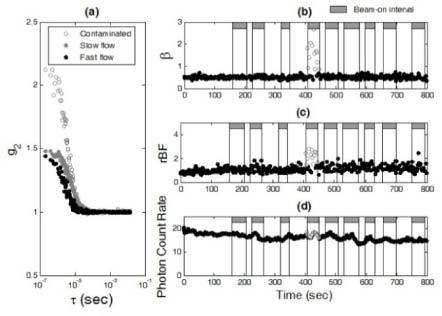
Fig. 3.

The in vivo measurement results for the investigation of optical measurement artifacts induced by scattered x rays. (a) Three autocorrelation curves (g2) were collected at the time points of 417 (contaminated), 99 (slow flow), and 724 (fast flow) (sec), respectively. Similar to the phantom test results (see Fig. 2a), scattered x rays contaminated the measured autocorrelation curves (empty circles). For the data without contaminations (solid circles), the decay rate of autocorrelation curves depended on the level of blood flow; g2 decayed faster when blood flow was faster (black solid circles). As expected, β (can be estimated using the measured g2 data at earliest τ) was independent of blood flow changes when there were no x-ray induced artifacts. (b) Scattered x rays created abnormal increases in β (>0.5, empty circles) depending on the direction/angle of radiation beam. (c) The x-ray beam introduced abnormal increases in blood flow (empty circles) derived from the autocorrelation curves. (d) The x-ray beam induced minor variations in detected photon count rate ( × 103 photons/s) (empty circles).
