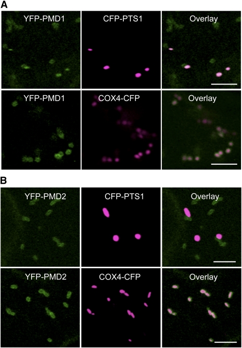
Figure 2.
Subcellular Localization of YFP-PMD1 and YFP-PMD2.
Confocal images are from leaf epidermal cells from transgenic plants expressing PMD1pro:YFP-PMD1 (A) or 35Spro:YFP-PMD2 (B) along with the peroxisomal marker CFP-PTS1 or the mitochondrial marker COX4-CFP. YFP signals are in green, and CFP signals are in magenta. Merged images show the colocalization of the YFP fusion protein to peroxisomes or mitochondria. Bars = 5 μm.

