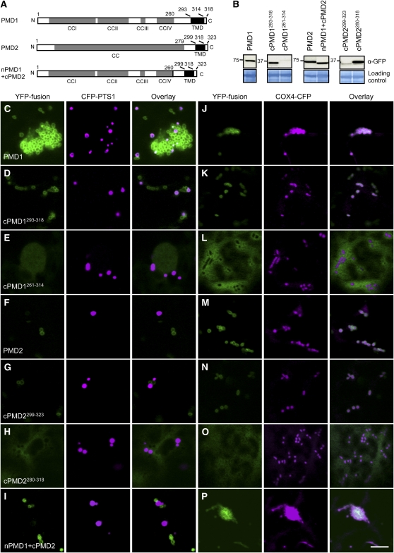
Figure 7.
Organelle Targeting Signals Reside in the C Terminus of PMD1 and PMD2.
(A) Schematics of PMD1, PMD2, and nPMD1+cPMD2 with the CC domains, TMD, and amino acids indicated. Despite sequence similarities between PMD1 and PMD2, PMD2 was annotated as having a single and long CC domain.
(B) Immunoblot analysis to detect the YFP-PMD variants expressed in tobacco cells. The large subunit of ribulose-1,5-bis-phosphate carboxylase/oxygenase was used as the loading control. Numbers to the left of each panel indicate molecular mass in kilodaltons.
(C) to (P) Subcellular targeting of the YFP-PMD1/PMD2 fusions. YFP signals are in green, and CFP signals are in magenta. To better illustrate colocalization between the YFP fusion and CFP-PTS1, images in (C) were taken from a region where peroxisome proliferation/aggregation was not so strong. Bar = 10 μm.

