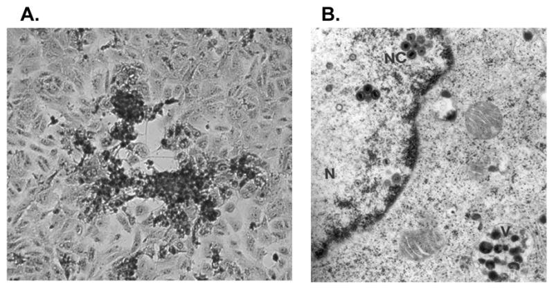
Figure 1.

SVV-infected Vero cells. (A) Cytopathic effect in a SVV-infected Vero cell monolayer (20X). (B) Electron microscopy of a SVV-infected Vero cell. SVV nucleocapsids (NC) within the cell nucleus (N) and degraded virions within cell vacuoles (V) are shown. Reprinted with permission by Springer Science and Business Media from Oakes and d’Offay in Virus Diseases in Laboratory and Captive Animals 1988; 163–174. Copyright Kluwer Academic/Plenum Publishers.
