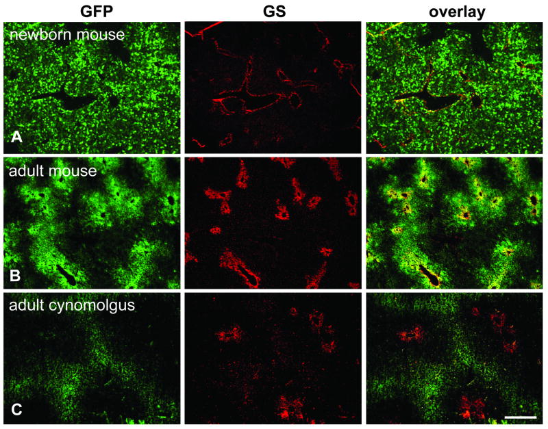
Fig. 2.
Immunofluorescence staining for GFP and GS on livers from animals that received self-complementary (sc) vectors expressing GFP. A. Newborn mouse (group 6) injected with 5×10¹⁰ GC of scAAV8.TBG.EGFP and analyzed seven days later. B. Adult mouse (group 4) treated with 3×10⁹ GC of the same vector seven days after injection. C. Adult cynomolgus macaque (24456) injected with 2×10¹² GC/kg of scAAV7 expressing GFP from a CB promoter. Scale bar: 400 μm.

