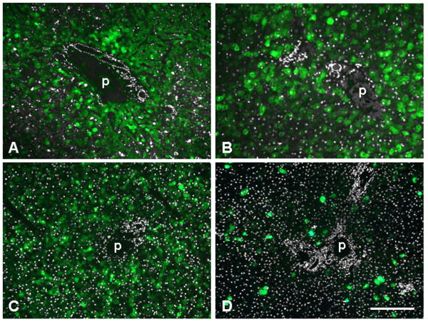
Fig. 5.
GFP expression in portal areas of liver from non-human primates after administration of 3×10¹² GC/kg of AAV8 expressing GFP from the TBG promoter. Shown is GFP fluorescence (green) overlaid with DAPI staining (white) to demonstrate bile duct and hepatic artery. A. Adult cynomolgus macaque C13991 seven days after injection. B. Adult rhesus macaque 607213 seven days after injection. C. Infant rhesus macaque N1, injected 1 week after birth and analyzed seven days later. D. Infant rhesus macaque N5, injected 1 week after birth and analyzed 35 days later. p indicates portal vein. Scale bar: 200 μm (A, C, D) and 130 μm (B).

