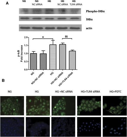
Figure 8.
Effect of TLR4 knockdown on HG-mediated IκB/NF-κB signaling in PTECs. (A) Western blot analysis of IκB phosphorylation. PTECs pretransfected with either TLR4 siRNA or negative control siRNA were treated with HG (30 mM) for 6 hours. The phosphorylation state of IκB was detected by immunoblotting against anti–phospho-IκB antibody. Levels of phosphorylation were normalized to actin. Results are means ± SD obtained from three independent experiments. *P < 0.05, versus PTECs cultured with NG media §§P<0.01 versus PTECs transfected with NC siRNA and cultured with HG media. A representative blot is shown at the top. (B) Study of subcellular translocation of NF-κB by immunofluorescence staining. PTECs with no knockdown, transfected with negative control siRNA or TLR4 siRNA, were incubated with HG (30 mM) for 8 hours and were stained by immunofluorescence for the p65 subunit of NF-κB (green, top panel) and for cell nuclei with 4′,6-diamidino-2-phenylindole (blue, bottom panel). PTECs treated with normal glucose (5.5 mM) and HG plus pyrrolidine dithiocarbamate (PDTC; an NF-κB translocation inhibitor) served as baseline and positive controls, respectively. NC siRNA, negative control siRNA; NG, normal glucose. Original magnification, ×400.

