Figure 1.
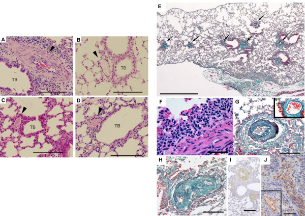
Histopathological features of the lung disease in B6.TgL mice. (A-D) Histopathological manifestation typically present in B6.TgL (A). No pathological manifestation observed in B6 (B), BALB.TgL (C), and BALB (D). The photograms indicated were taken from over 20-week aged male mouse. Arrow heads indicate pulmonary arteries. TB, terminal bronchiole. H&E staining. Scale bar = 100 μm. (E) Diffuse pathology present in B6.TgL. Masson's trichrome staining. Scale bar = 1 mm. (F) Perivascular lymphocytic infiltration in the affected lung. H&E staining. Scale bar = 50 μm. (G, H) Representative microscopic appearance in the affected arteries in the B6.TgL lung. An inset photogram in G represents the appearance of a normal pulmonary artery. Thickening of the intimal and, to a lesser extent, medial layers with marked intimal fibrosis is characteristic of the affected arteries. Masson's trichrome staining. Scale bar = 100 μm. I and J, expression of αSMA and CD31 (PECAM), respectively in the thickened arterial wall. The photogram in the inset of J represents a normal manifestation of unaffected artery. Immunohistochemical staining with hematoxylin counter-staining. Scale bar: in I, 200 μm; in J, 100 μm.

