Figure 3.
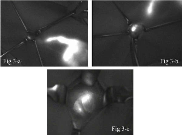
It is possible to see the fluorescent lymph vessels that run from the body surface to the axilla. Figure 3-a: A skin incision is made at the end of fluorescence emission that extends to the axilla, in order to observe the fluorescent lymph vessels embedded in the subcutaneous tissue. The fluorescent lymph nodes at the end of the lymph vessels are identified. Figure 3-b: After skin incision, the subcutaneous lymphatics were more clearly visible by fluorescence. Figure 3-c: The lymphatic channels and nodes that received ICG appeared as shining fluorescent streams and spots in the fluorescence image. The lymph node was dissected along with its surrounding fatty tissue.

