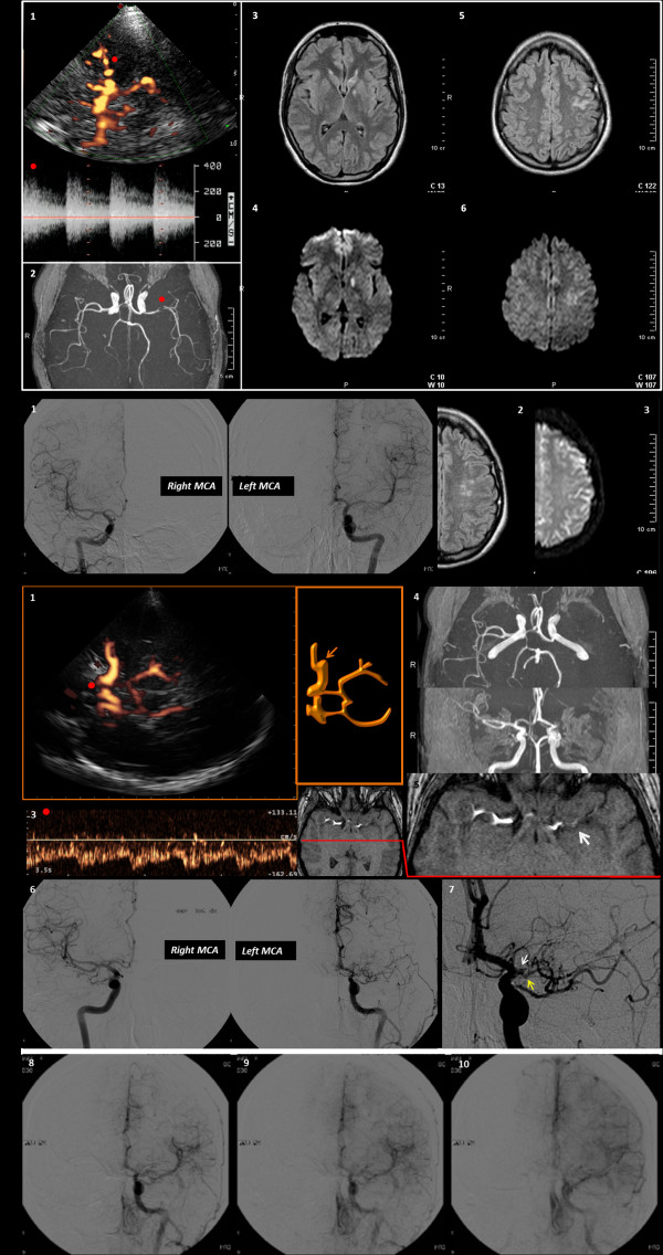
Figure 1.
Neuroimaging data of the patient. The neurosonological and neuroradiological findings are shown. Row A refers to the first occurrence of stroke. 1. TCCS image with contrast agent from the left temporal bone window in the mesencephalic axial plane (power mode). The red dot points to the left M1 MCA, with the corresponding flow spectrum. 2. MRA with TOF reconstruction: the red dot points to the left MCA stenosis. 3. T2-weighted MRI with the caudate head lesion 4. Corresponding positive DWI-MRI 5. T2-weighted MRI with cortical sulcal damage 6. Corresponding positive DWI-MRI. Row B shows: 1. DSA images of right and left MCA, with the confirmed mild left M1-M2 stenosis. 2. T2-weighted MRI of the left hemisphere with the signs of the previous infarction. 3. corresponding negative DWI-MRI. Row C refers to the third neuroradiological and neurosonological control examinations. 1. TCCS image with contrast agent from the left temporal bone window in the mesencephalic plane (Power mode), showing the left main stem MCA stop, and the early MCA branch. 2. Corresponding schematic drawing. 3. The Doppler waveform of the left A1 ACA is showed (the red dot points on the corresponding vessel segment in the Power-mode TCCS image), and the increased flow velocity suggests a condition of flow diversion for contributing to the reperfusion of MCA territory. 4. TOF MRA reconstruction with absent signal from left M1 MCA. 5. MRA source image with the proximal left M1 MCA branch, nearest to the origin of the vessel, coursing along the silvian fissure (white arrow); see for comparison the normal right MCA in the same figure. 6. DSA image, showing a good correspondence with the neusonological findings, comparing the right and the left MCA. 7. Zoomed detail of the very early left MCA branching (yellow arrow) on DSA, just before the MCA occlusion, nearest to the carotid bifurcation (white arrow).8. 8, 9, 10 Temporal sequence of selective DSA with contrast injection in the left. 9. ICA, showing the slow reperfusion of the distal MCA territories by the early. 10. MCA branch and the contribution of the anastomosis with distal branches of the left ACA..

