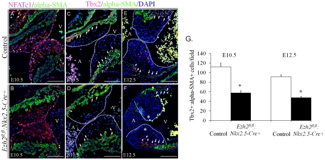
Figure 5. Decreased Tbx2 expression in the AV myocardium of Ezh2 mutant mice.
(A–B) Double immunofluorescence staining of the sagittal sections of E10.5 embryos with anti-NFATc1 antibody (red) and anti-alpha-SMA (green) revealed no change in the number of NFATc1+ endothelial cells in Ezh2-cKO hearts compared with that in littermate control hearts. (C–F) Double-immunofluorescence staining of the sagittal sections of E10.5–12.5 embryos with anti-Tbx2 antibody (red) and anti-alpha-SMA (green) revealed greatly reduced Tbx2+:α-SMA+ (arrows) primary myocardial cells of the atrioventricular canal in Ezh2fl/fl:Nkx2.5-cre+ hearts, compared with that of the littermate control hearts. Dotted lines outlined the valve primordia. The number of Tbx2+:α-SMA+ cells was counted in three randomly selected fields of Ezh2fl/fl:Nkx2.5-cre+ and control hearts of E10.5 or E12.5 embryos. Nuclei were counterstained with DAPI, as shown in blue. G shows the statistical analysis of Tbx2+:α-SMA+ cells between these two groups. Data are presented as mean ± SD. n = 3 for each group. *, p<0.05. Magnification, 20×. Scale bar: 100 µm.

