Fig. 3.
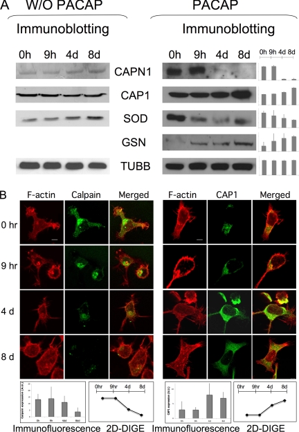
Validation of the DIGE/MS results by immunoblot and immunofluorescence analysis. A, Conventional 1D SDS-PAGE gels were run with proteins from CHRF cells treated with PACAP at several time points. Proteins were transferred onto nitrocellulose membranes and incubated with antibodies against some of the proteins identified in DIGE/MS analysis: calpain (CAPN1, 28kDa), adenylyl cyclase-associated protein 1 (CAP1, 52kDa), superoxide dismutase (SOD1, 16kDa) and gelsolin (GSN, 86kDa). Beta-tubulin (TUBB, 50kDa) was used as the internal loading control. The densitometric quantification of protein patterns showed similar trends with DIGE data for all proteins analyzed. Data obtained by performing the same experiment with vehicle PBS indicate that the change in protein expression observed after PACAP treatment are PACAP related and not due to CHRF cell growth. B, CHRF cells at the different points following PACAP treatment were plated on coverslips precoated with fibrinogen (100 μg/ml) 1 h at 37 °C. Cells were stained with rhodamin-phalloidin to visualize F-actin and after with Alexa468 anti-CAPN1 or -CAP1 antibody. The degrees of differential expression in the different time points for both DIGE (supplemental Table S1) and immunohistochemical analysis are highly consistent and are shown in the graphics as average of three experiment replicates ± standard deviation. Bar = 5 μm.

