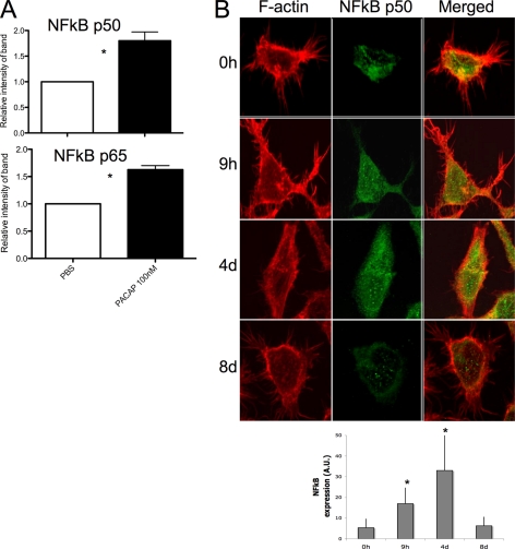
Fig. 6.
PACAP stimulates NF-κB signaling in CHRF cells. A, CHRF cells were stimulated for 1 h with 100 nm PACAP38 or vehicle PBS. Nuclear extracts were analyzed by immunoblotting for NF-κB subunits p50 and p65. Higher nuclear levels of p50 and p65 (p < 0.05) were observed when PACAP was added to the cells. B, CHRF cells at the different time points following PACAP treatment were plated on coverslips precoated with fibrinogen (100 μg/ml) for 1 h at 37 °C. Cells were stained with rhodamin-phalloidin to visualize F-actin and after with Alexa468 anti-NF-κB p50. The increased expression of NF-kB subunits after PACAP treatment were consistently detected by both immunoblot and immunofluorescence analysis. Figures show the average of three independent experiments + standard deviation. *p < 0.05.

