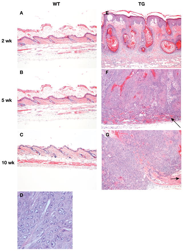
Figure 3. Histological appearance of lesions and tumors after DMBA.
Mice were topically treated with DMBA (400 μg/200 μl). At specified times after treatment, dorsal skin was removed, fixed in formalin, and stained with H&E and photographed. A–C, wild type epidermis (100×). D: Squamous cell carcinoma from BK5.EP1 mice (400×). E–G, lesions and tumors from BK5.EP1 mice.

