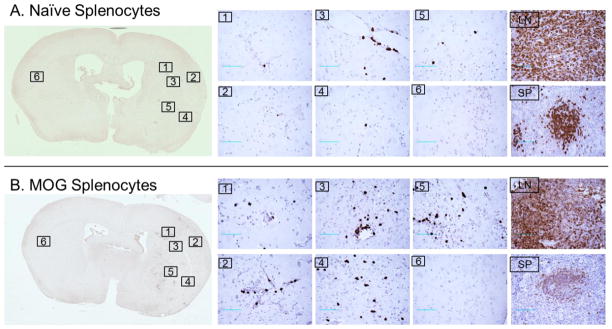
Figure 3.
Detection of GFP+ cells in brain, lymph node and spleen by immunohistochemistry after adoptive transfer of naive (A) or MOG stimulated (B) GFP+ splenocytes following 60min MCAO and 96h reperfusion. Sections are shown from Brain (with higher 40X magnifications shown from six comparable locations), lymph nodes (LN) and spleen (SP) from recipient mice after transfer of GFP+ splenocytes (Scale bars: 200 μm). Results represent one of three independent experiments that included five animals per group.

