
Figure 1.
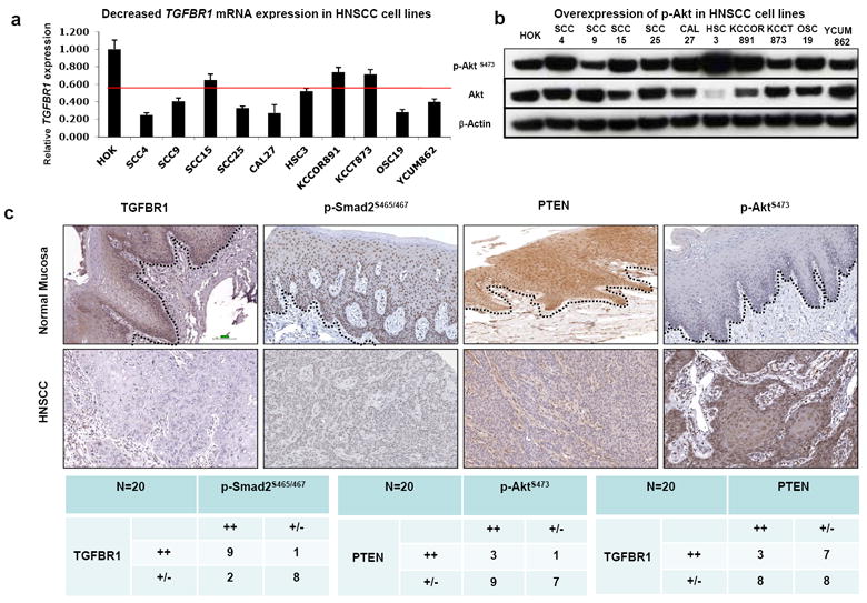
Loss of TGFBR1 and activation of PTEN/PI3K/Akt pathway in HNSCC samples. (a) TGFBR1 mRNA significantly reduced in HNSCC cell lines by qRT-PCR. Human oral keratinocytes (HOK) were used as normal control. (b) Western blot analysis demonstrates that p-Akt was overexpressed in most of the HNSCC cell lines. (c) Tissue array analysis revealed that TGFBR1, p-Smad2, and PTEN expression were reduced, while p-Akt was overexpressed in most human HNSCCs by IHC. The dotted lines delineate the adjacent epithelial compartment (magnification, 200X). Bar=100um.