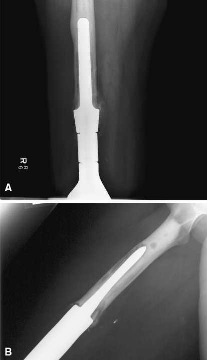
Fig. 2A–B.
(A) This radiographs shows an example of the large stem size relative to the diaphyseal width technique described in this article. It also shows cortical hypertrophy that can occur with transfer of the stress to the tip of the prosthesis. (B) The bottom radiograph is an example of the large bone:stem ratio technique with an undersized stem relative to the diaphyseal canal.

