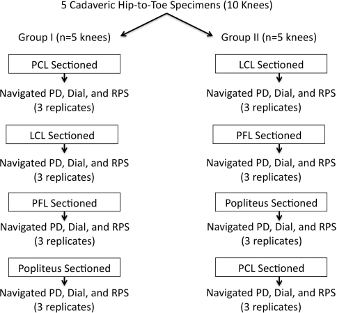
Fig. 1.
A flowchart shows the distribution of the 10 cadaveric hip-to-toe specimens utilized in this protocol. Five knees were allocated to each group (I and II). In Group I, the PCL was sectioned, followed by sequential sectioning of the structures of the PLC. In Group II, the structures of the PLC were sectioned followed by the PCL. Mechanical testing consisting of a posterior drawer (PD) test, external rotation dial tests at 30° and 90°, and RPS were performed for each condition.

