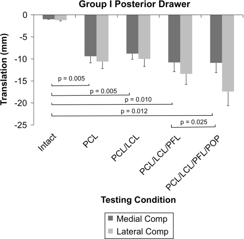
Fig. 5.
A graph shows medial and lateral compartment translations in response to a 65-N posterior drawer in Group I. Sectioning of the PCL resulted in an increase in posterior translation of the medial and lateral compartments. Further sectioning of the PLC structures did not result in an increase in medial compartment translation. Comp = compartment; POP = popliteus tendon.

