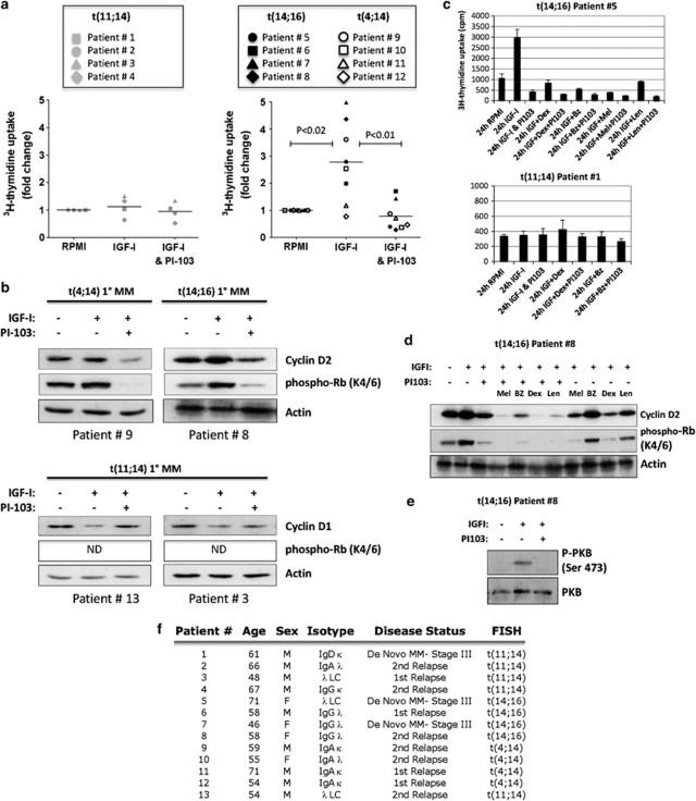
Figure 5.
Effect of IGF-I and PI-103 on cell cycle progression in primary MM cells. (a) Proliferation assays were performed on selected CD138+MM cells cultured for 24 h in either serum-free medium (RPMI-1640) alone or with 100 ng/ml IGF-I±1 μ PI-103, as indicated. Each data point represents the mean fold change in [3H]-thymidine uptake in triplicate wells for individual patients. The mean fold change in [3H]-thymidine uptake for each group of patients for each culture condition is denoted by a horizontal bar. Data from cyclin D1-expressing t(11;14) patients (n=4) are shown in the left-hand panel; those from cyclin D2-expressing t(4;14) and t(14;16) patients are in the right-hand panel. (b) Western blots of whole-cell lysates prepared from primary patient CD138+ cells treated as described above, were probed with antibodies against cell cycle control proteins, as indicated. Anti-actin is shown as a loading control. (c) The effect of PI-103 in combination with chemotherapeutic drugs on [3H]-thymidine uptake in two patients' CD138+ cells, treated as described. (d) The effect of PI-103 in combination with chemotherapeutic drugs on the expression of cell cycle proteins in lysates prepared from cells from patient no. 8 treated as described above. (e) Primary CD138+ cells were treated with 100 ng/ml IGF-I±1 μ PI-103 in serum-free RPMI-1640 medium for 15 min and analysed for PKB activation by western blotting of whole-cell lysates. Equivalent loading is demonstrated by total PKB expression. (f) Table showing details of patient samples used. BZ, bortezomib; Dex, dexamethasone; Len, lenalidomide; Mel, melphalan.

