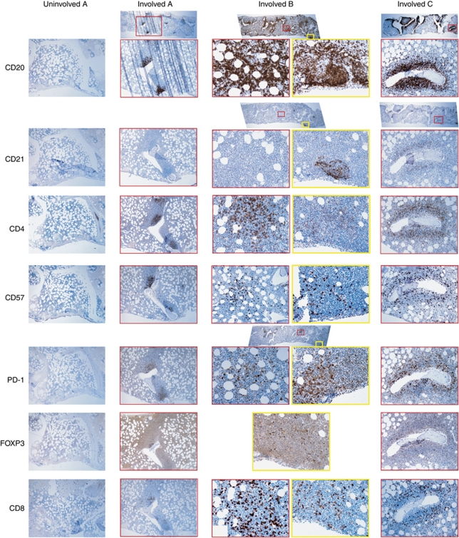
Figure 3.
Immunohistochemistry of bone marrows. All investigated bone marrows without follicular lymphoma involvement had the same appearance (left-most column, Uninvolved A), showing few and scattered cells positive for CD20, CD4, CD57, FOXP3 and CD8. There are no cells positive for CD21 or PD-1. This bone marrow is from the patient who later progressed to bone marrow involvement (Involved A), without any interceding therapy. The CD20 staining in the later biopsy shows a moderate infiltration of lymphoma cells. There are no accompanying CD21+ follicular dendritic cells. CD4+ and CD57+ T cells are mostly located in the infiltrates. PD-1+ T cells are exclusively found in the infiltrates and also FOXP3+ cells have homed there. There are some CD8+ cytotoxic T cells in the periphery of the infiltrates. Involved B is a bone marrow with heavy involvement of follicular lymphoma. Two involved areas (the red and yellow squares) have been examined in large magnification. CD21+ follicular dendritic cell networks are seen in one area, but not in the other, suggesting different stromal cells' support in the same specimen. The distributions of cells positive for CD4, CD57, PD-1 and CD8 seem to differ between the two areas. In both areas, FOXP3+ showed a perifollicular pattern. Involved C shows peritrabecular follicular lymphoma infiltrates. There is a CD21+ follicular dendritic cell network and an aggregation of CD4+, CD57+, PD-1+ and FOXP3+ T cells.

