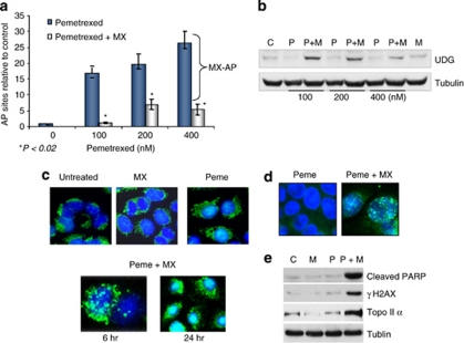
Figure 7.
MX-bound AP sites induced by the combination of pemetrexed and MX are lethal DNA lesions. (a) The dose-dependent relationship between the levels of AP sites and the concentrations of pemetrexed. The AP sites in DNA were measured using ARP reagent after H460 cells were treated with pemetrexed alone (0–400 nM) or in combination with MX (6 mM) for 24 h. MX-bound AP sites were determined by the differences between AP sites induced by pemetrexed alone and the combination of pemetrexed and MX. (b) UDG induction detected in cells treated with the pemetrexed alone and in combination with MX. Cells were collected and UDG protein was measured by western blotting analysis. (c) H460 cells grown on the coverslip were treated with pemetrexed alone (100 nM) and in combination with MX (6 mM) for 24 h and subjective to the fluorescent immunostaining. The signal of UDG protein (green) was significantly enhanced and localized in nucleus (blue) of cells treated with the combination of pemetrexed and MX. (d) γH2AX foci formation detected in H460 cells treated with pemetrexed (100 nM) alone and in combination with MX (6 mM) for 24 h; (e) Induction of the cleaved PARP and γH2AX proteins was detected by western blotting in cells with the same treatments

