Figure 2.
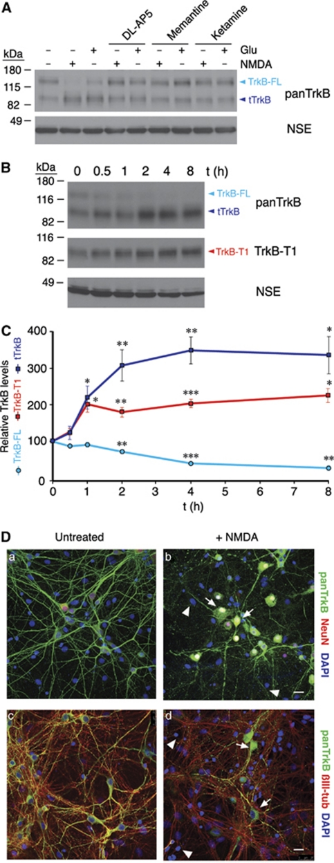
Neuronal-specific regulation of TrkB is induced by NMDAR overactivation in a cellular model of excitotoxicity. (A) Opposite regulation of the TrkB isoforms was induced by agonists of glutamate receptors and prevented by specific NMDAR antagonists. Primary cortical cultures (14 DIV) were treated with NMDA (100 μM) or glutamate (100 μM) with their co-agonist glycine (10 μM) for 2 h, with or without the antagonists DL-AP5 (200 μM), memantine (10 μM), or ketamine (500 μM). TrkB-FL and the other truncated isoforms (tTrkB) that are recognized by panTrkB are indicated. (B) Time course of the TrkB-FL decrease and the tTrkB or TrkB-T1 increase in cultures treated with NMDA for the indicated times. (C) Quantitation of TrkB dysregulation. Relative protein levels of TrkB-FL, tTrkB, or TrkB-T1 (mean±S.E.M., n=4 experiments) were established after normalization to NSE and comparison with the protein levels in untreated cells, which were arbitrarily assigned a value of 100%. The effect of NMDA was assessed using a Student's unpaired t-test (*P<0.05, **P<0.01, and ***P<0.001). (D) Double immunofluorescence of cultures stimulated with NMDA for 1 h using panTrkB or neuronal-specific antibodies demonstrated the neuronal specificity of TrkB expression (arrows) and the lack of TrkB staining in glial cells in the culture (arrowheads). Analysis with isoform-specific antibodies was not possible, because they produced very weak immunofluorescent signals. Representative confocal microscopy images corresponding to single sections are shown. The scale bars represent 10 μm

