Figure 4.
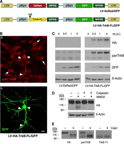
Characterization of TrkB-FL as a calpain substrate. (A) Schematic diagram of lentiviral vectors LV-DsRed/GFP (encoding DsRed and GFP fluorescent proteins) and LV-HA-TrkB-FL/GFP (coding for HA-TrkB-FL and GFP). The recombinant proteins are regulated by neuro-specific synapsin promoters (pSyn), and their sequences are followed by woodchuck hepatitis post-transcriptional regulatory element (WPRE) for the enhancement of expression. LTR, long terminal repeat. (B) Double immunostaining with panTrkB and GFP antibodies of cultures infected with a low multiplicity of LV-HA-TrkB-FL/GFP demonstrated increased TrkB levels in specific neurons co-expressing GFP (arrowheads) compared with endogenous TrkB in uninfected neurons (arrows). Confocal microscopy images correspond to single sections. The scale bars represent 10 μm. (C) Cultures infected with lentivirus LV-HA-TrkB-FL/GFP using increasing multiplicities of infection (m.o.i.) showed the expression of HA-TrkB-FL and the specific increase of TrkB-FL total levels compared with LV-DsRed/GFP-infected cells. (D) Processing of recombinant HA-TrkB-FL expressed in LV-HA-TrkB-FL/GFP-infected neurons (m.o.i.=2) is induced by 2 h of NMDA treatment and produces N-terminally HA-tagged fragments. Cleavage is blocked by pre-incubation for 1 h with the specific calpain-inhibitor calpeptin (10 μM). (E) In-vitro calpain processing of extracts prepared from neurons infected with LV-HA-TrkB-FL/GFP using purified calpain I (80 U/ml) directly established TrkB-FL as a calpain substrate, in contrast to TrkB-T1, and suggested that cleavage yields a truncated TrkB-FL that is similar to TrkB-T1. The additional HA-TrkB-FL fragment that was produced in vitro, with a mobility ∼110 kDa (left panel), may correspond to an intermediate product and was not observed in cultures subjected to excitotoxicity

