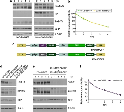
Figure 6.
Effect of increased TrkB-FL expression or TrkB-T1 interference during excitotoxicity. (a) Time course of NMDA treatment in cultures transduced with LV-HA-TrkB-FL/GFP (m.o.i.=2) demonstrated higher levels of total TrkB-FL compared with LV-DsRed/GFP-infected cells. (b) Relative neuronal viability (mean±S.E.M., n=6) of cells infected and NMDA treated as above. Using Student's unpaired t-test, no significant differences were found between cells infected with LV-HA-TrkB-FL/GFP or LV-DsRed/GFP. (c) Schematic diagram of lentiviral vectors for neuro-specific interference of the TrkB-T1 isoform. Pre-miRNAs specific for target sequences in TrkB-T1 mRNA, starting at nucleotide 2178 or 2310, were cloned under the control of pSyn to produce viruses LV-miT1(2178)/GFP and LV-miT1(2310)/GFP (herein, LV-miT1/GFP). Transcription of miRNAs was controlled by RNA polymerase II and was cell type-specific. Lentivirus LV-miC/GFP contains negative-control pre-miRNAs sequences. (d) Efficient and specific interference of basal TrkB-T1 expression was obtained by double infection with LV-miT1(2178)/GFP plus LV-miT1(2310)/GFP (m.o.i.=1 for each virus) or LV-miT1(2310)/GFP with LV-miC/GFP (m.o.i.=1 for each virus). No changes were observed for TrkB-FL or other analyzed proteins. (e) Double infection with LV-miT1(2178)/GFP and LV-miT1(2310)/GFP strongly interfered with the TrkB-T1 upregulation that was induced by NMDA compared with cultures infected with LV-miC/GFP (m.o.i.=2). (f) Relative neuronal viability (mean±S.E.M., n=6) of cells infected with LV-miT1(2178)/GFP and LV-miT1(2310)/GFP (m.o.i.=1 for each virus), or LV-miC/GFP (m.o.i.=2), treated or not treated with NMDA. Student's unpaired t-test revealed no significant differences in viability between cells infected with these viruses

