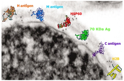
Figure 1.
Transmission electron micrograph of the surface of a H. capsulatum yeast cell with overlaying cartoons depicting the hypothetical structures of the H antigen, M antigen, heat shock protein 60, 70 kDa antigen, C antigen, and histone 2B. The antigen structures are based on molecular modeling as described in Guimaraes et al. (2008).

