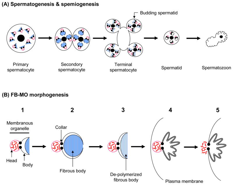
Fig. 3.
FB-MO morphogenesis during spermatogenesis and spermiogenesis. A: The cytological stages of spermatogenesis and spermiogenesis, and how they are coordinated with FB-MO morphogenesis. B: The stages of FB-MO morphogenesis. Red, wavy lines represent the contents of the MO vesicles, which are released into the extracellular space upon spermiogenesis. Blue lines show polymerized or de-polymerized MSP filaments. These figures are meant to show the fundamental process of FB-MO morphogenesis, so some details are simplified.

