
Figure 2.
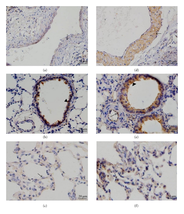
Immunohistochemical analysis of MIF in rat lungs. Immunostaining with an antibody to MIF of normoxic lungs ((a)–(c)) compared with lungs from hypoxic rats ((d)–(f)). The results showed no positive immunoreactivity for MIF in the pulmonary vasculature in control rats (arrow in (a) and (b)), but MIF stained smooth muscle cells of large pulmonary arteries (arrow in (d)), endothelial cells of small pulmonary arteries (arrow in (e)), and inflammatory cells around the alveoli (arrow in (f)) strongly in hypoxic lung sections. Arrows in (c) reveal normal alveoli. Arrowheads in (b) and (e) depict intense staining of bronchial epithelial cells.