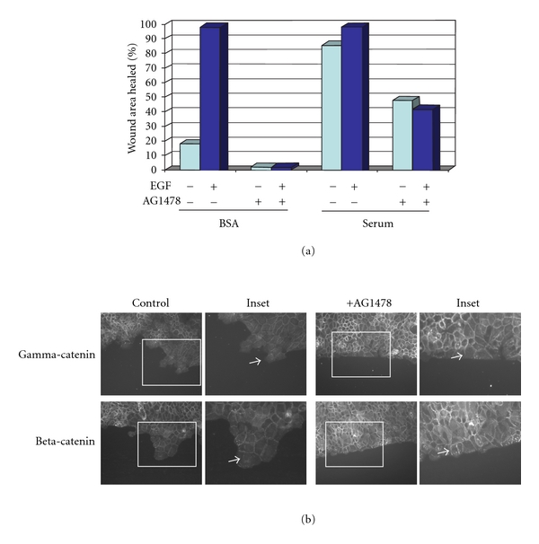
Figure 2.

Junctions at the wound margin are retained upon EGF receptor inhibition. Cells were grown to confluence, placed in serum-free medium overnight, and then a wound was introduced with a pipette tip. Cells were washed 3X with PBS, then placed in serum-free medium +/−20 nM EGF and +/−5 μM of the selective EGF receptor inhibitor AG1478 as indicated for 24 h. (a) The area of wound closure was measured using ImagePro software. (b) After wounding, cells were fixed and immunostained for either gamma-catenin (plakoglobin) or beta-catenin. Note the loss of border staining is limited to the wound edge (see arrows). Upon EGF receptor inhibition, junctional proteins are apparent at the wound edge (see arrowheads).
