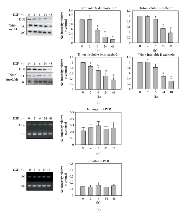
Figure 3.

EGF regulation of cadherin proteins in membrane and cytoskeleton-associated pools. Cells were grown to subconfluence, placed in serum-free medium overnight, and then treated with EGF for the indicated times. DG2: desmoglein-2, EC: E-cadherin, AC: alpha-catenin. (a) Sequential detergent extraction separates the triton soluble (membrane-associated) fraction, from the triton insoluble, (cytoskeletal, or intact junctional) fraction. Blots are representative of a minimum of three separate experiments. Bar graphs represent the densitometric quantification of each lane normalized to no treatment control, with asterisks indicating statistical significance. (P < 0.05) (b) Cells were treated with or without EGF for the indicated times, and mRNA level was measured by PCR. Bar graphs represent the densitometric quantification of bands normalized to no treatment control.
