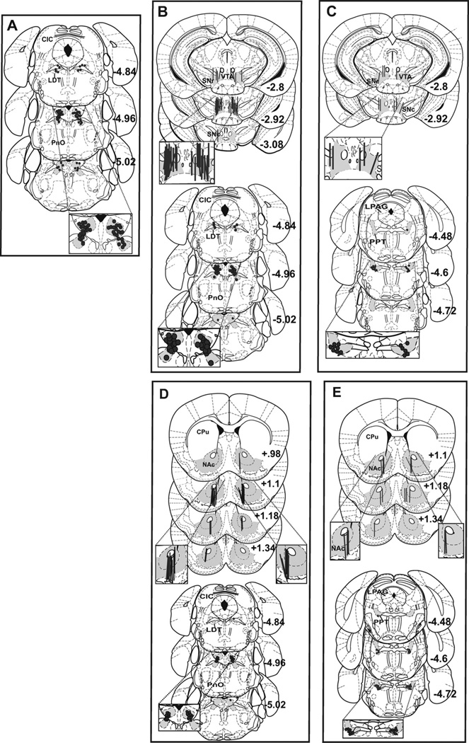
Fig. 1.
Microdialysis probe placements in the VTA and NAc and bilateral microinjection locations in the LDT and PPT are shaded and shown overlaid on plates taken from the atlas of Paxinos and Franklin [26]. Successful LDT microinjection targets for Experiment 1 are shown in panel A. Panels B and C show successful intra-VTA microdialysis probe placements for Experiments 2 and 3 (top) with the corresponding LDT (B, bottom) and PPT (C, bottom) microinjection locations. Panels D and E show successful intra-NAc microdialysis probe placements for Experiments 4 and 5 (top) with the corresponding LDT (D, bottom) and PPT (E, bottom) microinjection locations. Probe placements were counterbalanced for side and plates are labeled as mm from bregma. CIC: central nu of inferior colliculus; CPu: caudate putamen; LDT: laterodorsal tegmental nu; LPAG: lateral periaqueductal grey; NAc: nucleus accumbens; PnO: pontine reticular nu (oral part); PPT: pedunculopontine tegmental nu; SNc: substantia nigra, compact; SNr: substantia nigra, reticular; VTA: ventral tegmental area.

