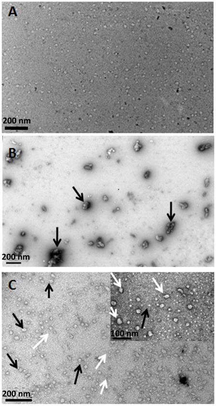
Fig. 3.
Representative TEM images of nanostructures detected in the 32-kDa enamelin and amelogenin solutions and their mixture, resulting from association of rP148 with enamelin in PBS buffer pH 7.4. (A) 32-kDa enamelin (0.032 mg/mL); (B) rP148 (0.2 mg/mL); (C) mixture of rP148 and enamelin at 10:1 ratio. Inset is a higher magnification.

