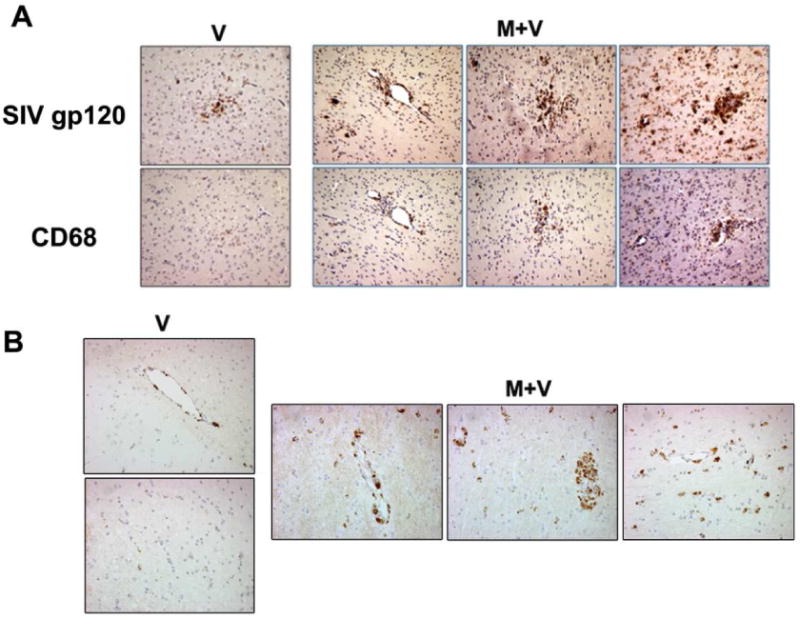
Figure 8. Localization of viral gp120 in the brain macrophages.

(A) Morphine treated macaques (M+V) showed more viral antigen and CD68+ macrophages in the basal ganglia compared to the SIV-treated (V) animal. The CD68 positive cells corresponded to regions that are also positive for the viral antigen gp120 indicating that macrophages constitute the majority of infected cells. (B) Representative image for CD68 staining in the basal ganglia section from V and M+V groups.
