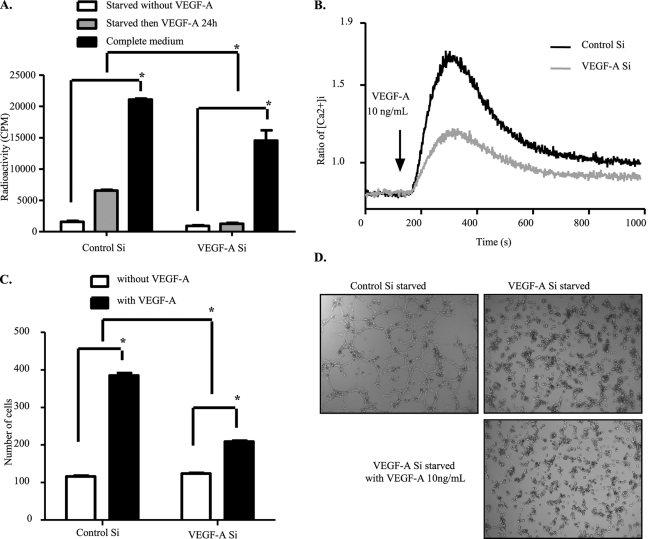
FIGURE 2.
Endothelial functions displayed by VEGFR-2 are impaired upon depletion of VEGF-A. A, shown is the reduced proliferation capacity of endogenous VEGF-A knockdown cells. HUVECs were transfected with control or VEGF-A siRNA for 48 h, and cell proliferation was then measured by [3H]thymidine incorporation assay. B, reduction in calcium release upon VEGF-A stimulation is shown. HUVECs were transfected with control or VEGF-A siRNA. Forty-eight hours later cells were digested and loaded with Fura-2. Intracellular calcium release upon VEGF-A stress was followed for 1000 s. C, cell migration capacity was affected by endogenous VEGF-A silencing. siRNA-treated cells were starved overnight. Forty-eight hours after transfection, cells were seeded into the upper chambers of Transwells and allowed to migrate into the bottom chambers containing VEGF-A165 (10 ng/ml) as the chemoattractant. Four hours later, cells were fixed and photographed for counting. D, tube formation was inhibited by depletion of endogenous VEGF-A. siRNA-transfected HUVECs were starved overnight, then seeded onto solidified Matrigel. VEGF-A165 (10 ng/ml) was then added into the culture medium, and tube formation was followed for 8 h. The data represented here are the average of three independent experiments. All bars represent means ± S.D. of three experiments. *, p < 0.05.

