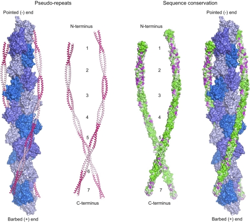
FIGURE 6.
Sequence conservation and Tm pseudo-repeats. Model of full-length Tm derived from the crystal structure of sm-Tmα81 and a published model of sm-Tmα (37) using MD simulations. Tm coiled coils, bound symmetrically along the two long pitch helices of the actin filament, are colored according to pseudo-repeat spacing (left) and amino acid conservation (right). Conservation decreases from green to magenta. Actin subunits are colored along the short pitch helix using three alternating shades of blue. Conservation scores were calculated with the program ConSurf (45) and displayed with the program PyMOL (DeLano Scientific LLC). The sequence alignment used to calculate the conservation scores is shown in supplemental Fig. 2.

