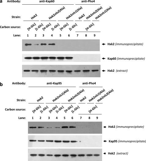
FIGURE 5.
Interaction of Kap60 and Kap95 with Hxk2. In vivo co-immunoprecipitation of Kap60 (a) and Kap95 (b) with Hxk2, Hxk2nls1(Ala), and Hxk2nls2(Ala). The wild-type, FMY304, and FMY305 strains were grown in YEPD media until an A600 nm of 0.8 was reached and then shifted to high (H-Glc) and low (L-Glc) glucose conditions for 1 h. The cell extracts were immunoprecipitated with a polyclonal anti-Kap60 or anti-Kap95 antibodies (lanes 1–6), or a polyclonal antibody to Pho4 (lanes 7–9). Immunoprecipitates were separated by 12% SDS-PAGE, and co-precipitated Hxk2 variants were visualized on a Western blot with monoclonal anti-Hxk2 antibody. The level of immunoprecipitated Kap60 or Kap95 in the blotted samples was determined by using anti-Kap60 and anti-Kap95 antibodies, respectively. The level of Hxk2 present in the different extracts used in Fig. 5, a and b, was determined by Western blot using anti-Hxk2 antibody. The Western blots shown are representative of results obtained from four independent experiments.

