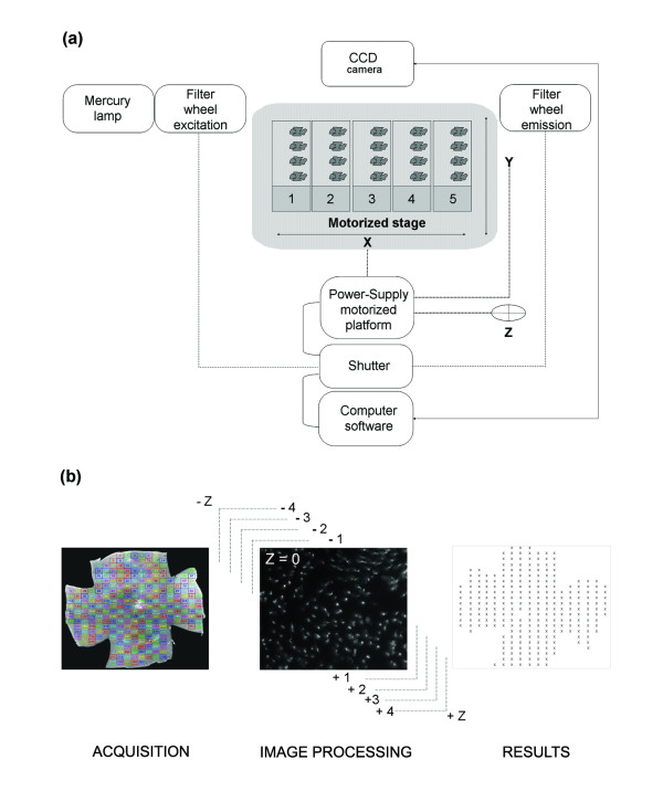
Figure 1.
℮-CONOME, the automated microscopy platform. (a) Connections between the different pieces of equipment. The motorized stage is made for five slides with a maximum of four retinas per slide. (b) Visualization of the grid used for acquisition and counting (left). Representative image of an acquisition for one field and the nine Z-planes (centre), the illustration corresponds to the best focus Z = 0 as indicated. Schematic representation of the results of global automated counting in an Excel sheet (right). The color code is providing an easy visualization of the fields of the grid during acquisition.

