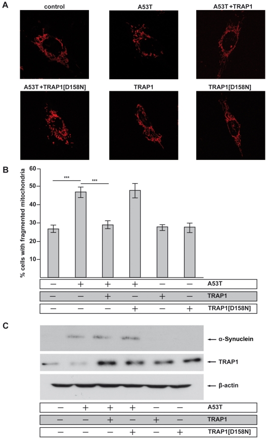
Figure 6. Inhibition of mitochondrial fusion by [A53T]α-Synuclein is rescued by TRAP1.
SH-SY5Y cells were co-transfected with the indicated constructs and mito-DsRed to visualize mitochondria. The mitochondrial morphology of transfected cells was analyzed by fluorescence microscopy. (A) Confocal images of representative cells displaying either an intact tubular mitochondrial network (control) or a fragmentation of this network (A53T). Co-expression of TRAP1[WT] prevented [A53T]α-Synuclein-induced mitochondrial fragmentation, whereas the TRAP1[D158N] mutant did not show rescue activity. Overexpression of either wild type or mutant TRAP1 alone did not influence mitochondrial morphology under steady state conditions. (B) For quantification, the mitochondrial morphology of at least 300 transfected cells per coverslip was determined in a blinded manner. Quantifications were based on triplicates of at least three independent experiments. Shown is the percentage of cells with fragmented mitochondria. (C) Expression levels of [A53T]α-Synuclein and TRAP1 were analyzed by Western blotting. β-Actin served as a loading control. ***p<0.001 (ANOVA).

