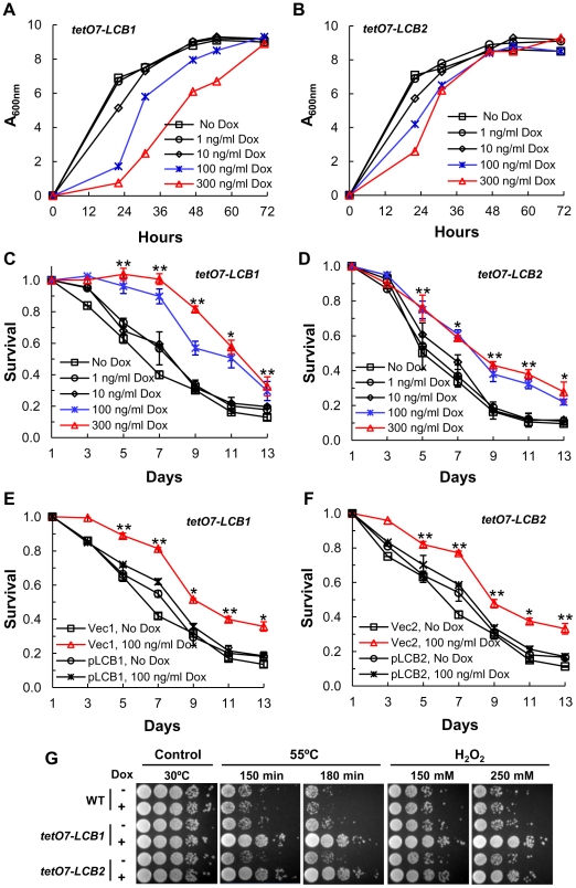
Figure 2. Down-regulating sphingolipid synthesis increases CLS.
(A) Growth rate of tetO7-LCB1 cells (RCD956) treated with Dox to down-regulate expression of the tetO7 promoter and reduce the rate of sphingolipid synthesis. (B) Growth rate of tetO7-LCB2 cells (RCD957) +/− Dox treatment. (C) CLS of tetO7-LCB1 cells +/− Dox treatment. Data show the mean ± SEM of surviving cells (* p<0.05, ** p<0.01, No Dox vs 100 ng/ml Dox). (D) CLS of tetO7-LCB2 cells +/− Dox treatment. Assays are similar to those presented in (C). (E) Verification that Dox-treatment extends CLS by reducing expression of the tetO7-LCB1 target gene. CLS of tetO7-LCB1 cells transformed with an empty vector (Vec1 = pRS315ADE2) or a vector carrying LCB1 (pLCB1 = pRS315ADE2-LCB1-5) is shown. Data represent the mean ± SEM of surviving cells (* p<0.05, ** p<0.01, for Vec1, 100 ng/ml Dox vs pLCB1, 100 ng/ml Dox). (F) Control experiment showing that Dox-treatment extends CLS by reducing expression of the tetO7-LCB2 gene. CLS of tetO7-LCB2 cells transformed with a vector carrying LCB2 (pLCB2 = pRS315-LCB2-B7) or with an empty vector (Vec2 = pRS315) is shown. Data represent the mean ± SEM of surviving cells (* p<0.05, ** p<0.01, for Vec2, 100 ng/ml Dox vs pLCB2, 100 ng/ml Dox). (G) Resistance of Dox-treated WT (R1158), tetO7-LCB1 (RCD956) and tetO7-LCB2 (RCD957) cells to heat (55°C) or hydrogen peroxide (H2O2) stress. Photographs show a ten-fold dilution series of cells (from left to right).

