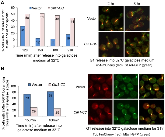
Figure 3. Overexpression of CIK1-CC leads to defects in chromosome bipolar attachment.
A. G1-arrested cdc13-1 CEN4-GFP TUB1-mCherry cells with a vector or a PGALCIK1-CC plasmid were released into galactose medium and incubated at 32°C. Cells were collected at the indicated time points and fixed for the examination of fluorescence signals. The relative localization of CEN4-GFP in cells with a normal looking metaphase spindle was determined. Of the cells with a metaphase spindle, the percentage of cells with a single GFP dot close to one spindle end was calculated and the result is shown in the left panel (n>100). The relative localization of CEN4-GFP signals to the spindle in some representative cells is shown in the right panel. B. cdc13-1 MTW1-GFP TUB1-mCherry cells with a vector or a PGALCIK1-CC plasmid were released into galactose medium and incubated at 32°C. Cells were collected at the indicated time points. Among the cells with a metaphase spindle, the percentage of cells with two clear GFP foci was counted and the result is shown in the left panel (n>100). The spindle morphology and Mtw1-GFP distribution in some representative cells are shown in the right panel.

