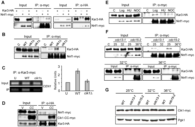
Figure 6. Kar3–kinetochore interaction is Cik1-dependent and cell cycle–regulated.
A. Kar3 interacts with Nnf1 in vivo. KAR3-3HA NNF1-13myc cells in mid-log phase were collected to prepare cells extracts. The extracts were immunoprecipitated with either anti-HA or anti-myc antibody. The protein levels in the whole cell extracts and precipitates are shown after Western blotting with anti-HA and anti-myc antibodies. B. The interaction of Kar3 with Nnf1 depends on Cik1. WT, cik1Δ, and vik1Δ cells carrying KAR3-3HA NNF1-13myc were grown to mid-log phase. The extracts were immunoprecipitated with anti-myc antibody. The protein levels of Kar3-3HA and Nnf1-13myc are shown after Western blotting. C. cik1Δ mutant cells show deceased Kar3-centromere interaction. KAR3-13myc and cik1Δ KAR3-13myc cells in log phase were collected for ChIP assay with anti-myc antibody. The PCR products with primers specific for CEN1 are shown in the left panel and the quantified data from three repeats are shown in the right. “U” is an untagged strain used as a control. D. Overexpression of CIK1-CC disrupts Kar3-Nnf1 interaction. KAR3-3HA NNF1-13myc cells with a vector (V) or a PGALCIK1-CC plasmid (CC) were incubated in galactose medium for 3 hrs. The protein samples were prepared and the Kar3-Nnf1 interaction was analyzed as described in A. E. Kar3 interacts with Nnf1 in HU- and nocodazole-arrested cells. KAR3-3HA NNF1-13myc cells in mid-log phase were divided into three portions as untreated (Log), HU (200 mM)- and nocodazole (NOC, 20 µg/ml) - treated samples. After 3 hr treatment, the samples were analyzed for Kar3-Nnf1 interaction as described. KAR3-3HA cells was used as a negative control and marked as ‘C’. F. Kar3 dissociates from Nnf1 in cdc13-1 and cdc15-2 arrested cells. cdc13-1 and cdc15-2 cells with KAR3-3HA and NNF1-13myc were grown to mid-log phase at 25°C. Then temperature was shifted to 32°C (for cdc13-1) and 36°C (for cdc15-2). After 2 hr incubation, the cells were collected and the protein samples were prepared and analyzed for Kar3-Nnf1 interaction. This interaction in cdc13-1 and cdc15-2 mutant cells incubated at permissive and non-permissive temperatures is shown in the top panel. The Kar3-Nnf1 interaction in WT cells after incubation at 32°C and 36°C for 2 hr is shown at the bottom panel. G. The Cik1 protein levels at different cell cycle stages. WT, cdc13-1, and cdc15-2 cells with CIK1-13myc were grown to mid-log phase at 25°C, and then shifted or 32°C (for cdc13-1) and 36°C (for cdc15-2) for 2 hrs. The cells were harvested to prepare protein samples. Cik1 protein levels are shown after Western blotting. Pgk1 proteins level was used as a loading control.

