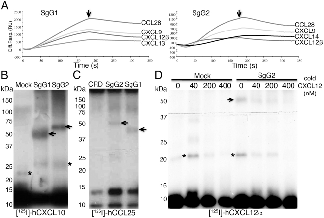
Figure 2. HSV-1 and HSV-2 gGs bind chemokines.
(A) Sensorgrams depicting the interaction between chemokines and SgG1 (left) or SgG2 (right). The indicated chemokines were injected at a 100 nM concentration. The arrow indicates the end of injection. All curves were analyzed with the BiaEvaluation software and represent the interaction of the chemokine after subtraction of the blank curve. Only 4 out of 11–12 positive interactions are shown. Abbreviations: Diff. Resp., Differential response; R.U., response units; s, seconds (B, C) Crosslinking assays showing the interaction of HSV-SgGs with [125I]-hCXCL10 (B) and [125I]-hCCL25 (C). Recombinant purified HSV-SgGs were incubated with iodinated chemokine and crosslinked with EGS (for [125I]-hCCL25) or BS3 (for [125I]-hCXCL10). The samples were resolved by SDS-PAGE, fixed and visualized by autoradiography. (D) Crosslinking assay between [125I]-hCXCL12α and SgG2 in the presence of increasing concentrations of cold hCXCL12α. Molecular masses are indicated in kDa. SgG-chemokine complexes are indicated with arrows and crosslinked chemokine dimers are marked with asterisks. Abbreviations: CRD, CrmB-cysteine rich domain.

