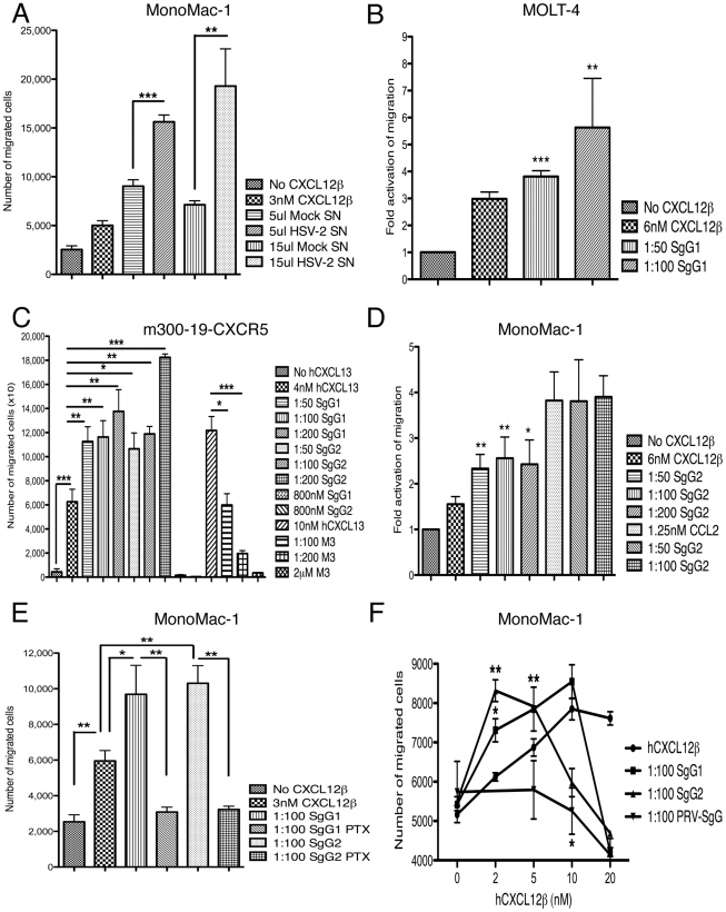
Figure 5. HSV SgGs enhance chemokine-mediated cell migration.
MonoMac-1 cells (A, D, E, F), MOLT-4 (B), m300-19-hCXCR5 (C) cells were incubated with the specified chemokine in Transwell plates. The effect of mock- or HSV-2-infected supernatant (Mock SN or HSV-2 SN, respectively) (A), purified SgG1 (B, C, E, F), SgG2 (C–F), M3 (C) and PRV-SgG (F) was analyzed. The number of migrated cells or the fold activation of migration is depicted. (C) SgG1 or SgG2 require the presence of the chemokine to enhance migration since addition of either of them without chemokine did not have any effect on chemotaxis. (D) Binding of HSV SgGs to the chemokine is necessary for the enhancement in chemotaxis. Representation of the fold activation of migration observed when cells were incubated with either hCXCL12β or hCCL2 in the absence or presence of increasing concentrations of HSV-2 gGs. (E) Addition of pertussis toxin (PTX) inhibits SgG-mediated enhancement of chemotaxis. Graph showing the effect of PTX addition on HSV SgGs enhancement of hCXCL12β-mediated chemotaxis. The number of migrated MonoMac-1 cells is represented. (F) HSV SgGs displace the hCXCL12β chemotactic curve towards lower concentrations of chemokine. MonoMac-1 cells were incubated with increasing concentrations of hCXCL12β in the absence or presence of a 1∶100 molar ratio of HSV SgGs or PRV-SgG. (A–F) Error bars indicate standard deviation values obtained from triplicate samples (A, C, E, F). One representative experiment of at least three is shown. In B and D, error bars represent the standard deviation in the fold activation obtained using three independent experiments performed in duplicate. *P<0.05; **P<0.01; ***P<0.001.

