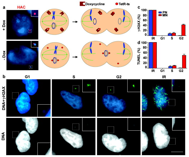
Figure 2. DNA breaks in a human artificial chromosome targeted to a micronucleus.
a, (Right) Schematic. (Left) Fluorescence in situ hybridisation images of HAC (red) in a primary nucleus (+Dox) or MN (-Dox). b, Images of micronucleated cells as in Fig. 1b (enlarged and brightened in insets). Scale bars, 10 μM c-d, Percentage of primary nuclei (blue bars) and centric MN (red bars) with (c) γ-H2AX foci and (d) TUNEL labelling. (3 experiments, n=100). Errors bars, s.e.m.

