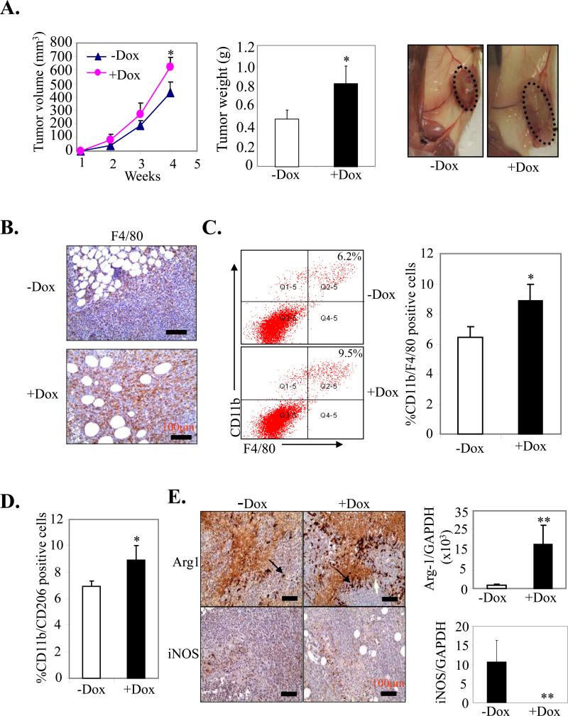
Figure 3. Effect of mS100a7a15 on tumor growth in orthotopic syngeneic model.
(A, left) MVT-1 cells were injected into the MG of the MMTV-mS100a7a15 mice (n=5) and tumor volume was measured every week. (A, middle) After 28 days, the tumors were excised from mice and weighed. (A, right) Representative photograph of mice showing tumors dissected from different experimental groups. (B) MVT-1 cell line derived tumors from +Dox and –Dox MMTV-mS100a7a15 mice were subjected to IHC staining for macrophage marker, F4/80. (C) CD11b+F4/80+ cells and (D) CD11b+CD206+ were quantified by flow cytometry in disaggregated MVT1 primary tumors harvested 28 d after implantation from +Dox and – MMTV-mS100a7a15 mice. (E, right) IHC of Arginase-1 (Arg-1) and iNOS. (E, left) Expression of Arg-1 and iNOS by q-PCR. Data represent the mean ± SD of three independent experiments. *, p<0.05 and **, p<0.01.

