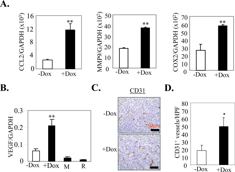
Figure 4. Effect of mS100a7a15 expression on pro-metastatic and angiogenic markers.
(A) Gene expression was quantified by q-PCR in mammary tumors from +Dox and –Dox MMTV-mS100a7a15 mice (n=5). (B) VEGF expression in +Dox and –Dox MMTV-mS100a7a15 mice and MVT-1 (M) or RAW264.7 (R) cell lines. (C) Representative IHC with endothelial marker CD31 antibody to assess the number of blood vessels in tumors from Dox treated compared to untreated mice. (D) Bars represent the mean ± SD of the number of CD31+ blood vessels shown in C counted in five random high power fields (HPF, 20X) per tissue section (n=5). *, p<0.05 and **, p<0.01.

