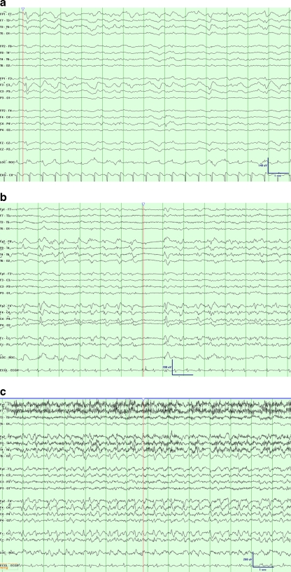
Fig. 1.
Examples of common electrographic patterns, displayed at 30 mm/s time base, 7 uV/mm sensitivity, 1-Hz low frequency filter, and 70-Hz high frequency filter. (a) A patient with recent subarachnoid hemorrhage following anterior communicating artery aneurysm clipping. Electroencephalogram (EEG) monitoring revealed frequent runs of left anterior lateralized rhythmic delta activity with admixed sharp components (LA-LRDA + s) at 1-1.5 Hz; time marker denotes additional epileptiform sharp components. (b) A patient with a newly diagnosed right temporal brain mass, presumed metastatic from hepatocellular carcinoma. The routine EEG showed right lateralized periodic discharges with admixed fast activity at approximately 1.5 Hz; time marker notes an associated period of relative background suppression. (c) The same patient depicted in (b), but with a subsequent period of chewing movements (note movement artifact), increased bilateral representation of the activity, and increased frequency. This discrete period of evolution is consistent with a seizure with a subtle clinical correlation. Shortly after EEG placement, the patient was found to be having seizures on an average of every 15 minutes

