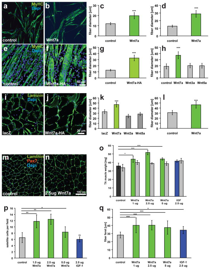
Figure 1.
Wnt7a induces hypertrophy in differentiated myotubes and myofibres. (a, b) Primary myoblasts derived from satellite cells were differentiated for 5 days in medium containing 50 ng/ml Wnt7a recombinant protein or BSA as a control. Staining for myosin heavy chain (MyHC, in green) marks differentiated cells. (c) Quantification of the fibre diameter as in a, b. n=3 (d) Quantification of the fibre diameter of C2C12 cells treated with 50 ng/ml recombinant Wnt7a protein. The Wnt7a recombinant protein was applied at day 3 of differentiation when the majority of the cells are already differentiated. n=3 (e,f) C2C12 cells were stably transfected with a CMV-Wnt7a-HA expression plasmid and differentiated for 5 days. (g) Quantification of the fibre diameter as in e,f. n=3 (h) Application of different recombinant Wnt proteins (50 ng/ml each) revealed that induction of hypertrophy is a Wnt7a specific phenomenon. n=3 (i,j) Electroporation of the CMV-Wnt7a-HA expression plasmid (40 μg) into the tibialis anterior muscle of adult mice resulted in increased fibre diameters 2 weeks after electroporation compared to a control (CMV-lacZ) plasmid. (k) Quantification of the fibre diameter of TA muscles electroporated with expression plasmids for Wnt3a-HA, Wnt5a-HA, Wnt7a-HA or a lacZ control plasmid (40 ug plasmid each). n=4 (l) Quantification of a single injection of recombinant Wnt7a protein (2.5 ug) into the tibialis anterior muscle of adult mice, mice were sacrificed two weeks after injection. n=4 (m,n) representative images of immunostained sections of TA muscle showing Pax7- (red) and laminin-staining (green). Nuclei were counterstained with Dapi (blue) (o) Intramuscular injection of recombinant Wnt7a protein into the tibialis anterior (TA) muscle resulted in a significant increase in the muscle weight, without affecting the weight of the contralateral muscle three weeks after injection. Injection of long-IGF stimulated an equal increase in both injected and contralateral TA muscle. n=4 (p) Wnt7a stimulated an expansion in the satellite cell pool whereas IGF did not. n=4 (q) Wnt7a and IGF injection resulted in a significant increase in fibre calibre. Grey bars indicate the contralateral muscle.. n=4, *p<0.01, **p<0.001, *** p<0.0001. Error bars represent SEM.

