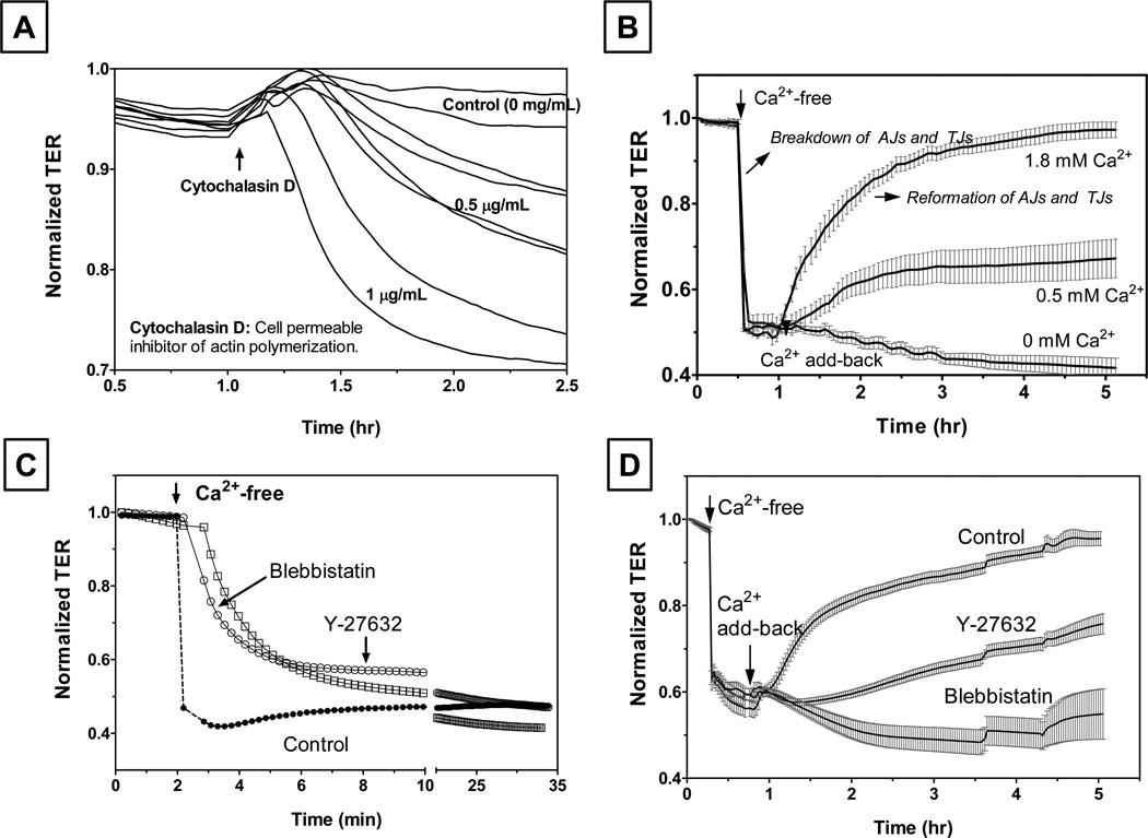
Figure 4. Reformation of TJs and AJs in bovine corneal endothelial monolayers assessed by trans-endothelial electrical resistance (TER).
Panel A: Loss of barrier integrity in response to cytochalasin D. At the up-arrow shown at about 1 hr after reaching the steady state, cells were exposed to cytochalasin D. As expected, cytochalasin D led to a decrease in the normalized resistance in a dose-dependent manner. Panel B: Breakdown and reassembly of apical junctions during Ca2+ Switch: When cells were exposed to Ca2+-free Ringers (with 2 mM EGTA), a precipitous fall in TER consistent with the breakdown of AJs is noticed. Ca2+ add-back led to a gradual recovery of TER. The rate and extent of recovery was dependent on the level of extracellular Ca2+. Panel C: Disassembly of AJs is opposed by inhibition of actomyosin contraction. Cells, pretreated with Y-27632 (Rho kinase inhibitor) or blebbistatin (Myosin II ATPase inhibitor) for 10 min, were exposed Ca2+-free Ringers. The rate of decrease in TER was smaller compared to the rate observed in the absence of Y-27632 and blebbistatin. Panel D: Reformation of AJs and TJs is also opposed by loss of actomyosin contraction. When cells were pre-exposed to Y-27632 or blebbistatin, recovery of TER in response to Ca2+ addition was inhibited. These results, adapted from our previous report (Ramachandran and Srinivas, 2009), suggest a role for actomyosin contraction in the disassembly and reformation of tight junctions.

